
| Properties | Details |
|---|---|
| SFDA Classification | Class IIb |
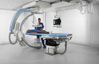
| Purpose of Use | Fluoroscopy is used in a variety of examinations and procedures. It can be used to diagnose or treat patients (in addition to using other tools). Some examples are: Barium X-ray: Fluoroscopy is used on its own and allows the doctor to see the movement of the intestines as the barium moves through it and allows the doctor to place the patient on intramural imaging. Cardiac catheterization: Fluoroscopy is used as an adjunct to enable the doctor to see the blood flow through the coronary arteries to assess the presence of arterial blockages. To insert the catheter into the vein, fluoroscopy helps the doctor guide the catheter to a specific location within the body. Image-guided injection of anesthetics into the joints or spine. Placing devices inside the body, such as stents, to open narrowed or blocked blood vessels. Angiograms (to visualize blood vessels and organs) Identifying a foreign body in the body. |
| Work Location & End-User | Place of work: Radiology department End User: Radiologist |
| Parts & Accessories | • X-ray generator • X-ray tube • candidate • patient table • Network • Image Intensifier • Video camera • Monitor 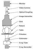 |
| Operation | • Take off clothing or jewelry and wear a medical gown. • Contrast material or X-ray dye may be given by swallowing (syrup or pills), or into an IV line in your hand or arm, depending on the type of procedure being performed. • You will be placed on an X-ray table. Depending on the type of procedure, you may be asked to move into different positions to take pictures of the body part from different angles. • For procedures that require catheter insertion, a needle may be inserted into the groin, elbow, or any other part. • The X-ray tube is directed to the part of the body to be examined. • A beam of X-rays will be generated from the X-ray generator and will pass through the body part. • The image will be transmitted to a screen so that the movement of the body part can be seen in detail. • After the procedure is over, if a catheter has been placed it will be removed. |
| Common Problems | • Equipment has been calibrated incorrectly. • operator error. • No radioactive emission. • Defective sockets or plugs or cables connection. • Safety lock prevents operation of equipment. • Electrical or electronic malfunctions. • High voltage cable or x-ray tube failure. • - Mechanical problems. • High pressure problems. • A small black spot on the X-ray angle due to damage to the cassette or exposure of the film to light. In this case, replace the damaged cassette. • White spots on the image due to screen scratches. •Dark marks or excessive brightness due to static electricity. In this case, remove the film from the cassette. |
| Manufacturers | • Siemens Healthineers • Philips • GE Healthcare |
| Sources | • Saudi Food and Drug Authority • US Food and Drug Administration • Johns Hopkins Medicine • RadioGraphics • Nucleus Medical Media • University Hospitals Birmingham NHS • MedlinePlus • Medbox • Dentaltix • Siemens Healthineers • Philips • General Electric Healthcare |