
| Properties | Details |
|---|---|
| SFDA Classification | Class IIa |
| Purpose of Use | to visualize internal areas of the body which cannot be seen by the naked eye and to diagnose or treat certain diseases with low-risk minimally invasive procedure that requires only small incisions.
Laparoscopic surgery can be used for various procedures which include: 1. Cholecystectomy - Removal of the gallbladder 2. Removal of patches of endometriosis 3. Treatment of ectopic pregnancy 4. Biopsy 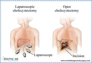 |
| Work Location & End-User | Work location: It is used in one-day surgery clinics and operating rooms End User: The doctor or specialist surgeon. |
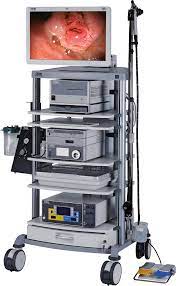
| Parts & Accessories | Basic devices supporting the laparoscope: 1. Light source with an intensity that can be controlled provide high intensity xenon, mercury and halogen lamp that delivered by fiberoptic bundle. 2. Video processor: receiving and processing signals from the camera 3. Monitoring screen: Show live video, patient information, control settings and save data 4. Insuffaltor: lifting the abdominal wall by insufflation with CO2 gas. 5. Suction-irrigation system: flushing the abdominal cavity and cleaning during laparoscopy. 6. Trolley The main parts of the laparoscope 1. Camera Connector: Connect the camera to the video processor 2. Objective lens 3. Light cable connection: provide different field of view by rotate the light cable. 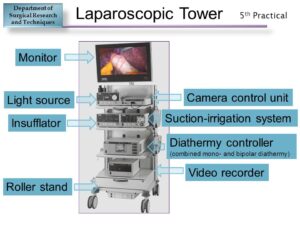 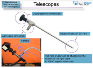 |
| Operation | 1. The surgeon uses the trocar to make multiple small holes that help insert the endoscopic instruments during surgery 2. Then the surgeon uses a carbon dioxide inflator to raise the abdominal wall, giving more space to work 3. Insert laparoscope to see inside the body and it work like endoscope: 1. The light source illuminates the area through the optical fiber 2. The objective lens of the CCD camera (Charge Coupled Device) collects the reflected light as signals and sends them to the processor. 3. The processor reads the signals, processes them and displays them as a video on the screen. 4. A suction tube is required to clean the area from excess liquid 5. Use other instruments for treatment or biopsy. 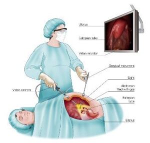 |
| Common Problems | • Refraction of the hard tube laparoscope • Refraction of some optical fibers, which causes a decrease in the efficiency of light transmission • Failure to distinguish between types of cameras leads to their disabling due to misuse. |
| Manufacturers | • Karl Storz • Stryker • Olympus |
| Sources | • Saudi Food and Drug Authority • DifferenceBetween.net • Patient.info • Department of Surgical Research and Techniques • World Laparoscopy Hospital • Etherealmind • Karl Storz • Stryker • Olympus |