
| Properties | Details |
|---|---|
| SFDA Classification | Class IIb |
| Purpose of Use | Mammography is used to detect breast cancer at an early stage by looking for signs that appear on X-ray images. This can lead to earlier treatment, provide a range of treatment options, and a better chance of survival. Barium X-ray: Fluoroscopy is used on its own and allows the doctor to see the movement of the bowel as the barium moves through it and allows the doctor to place the patient on intramural imaging. Cardiac catheterization: Fluoroscopy is used as an adjunct to enable the doctor to see the blood flow through the coronary arteries to assess the presence of arterial blockages. To insert the catheter into the vein, fluoroscopy helps the doctor guide the catheter to a specific location within the body. Image-guided injection of anesthetics into the joints or spine. Placing devices inside the body, such as stents, to open narrowed or blocked blood vessels. Angiograms (to visualize blood vessels and organs) Identifying a foreign body in the body. |
| Work Location & End-User | Place of work: Radiology department End User: Radiologist |
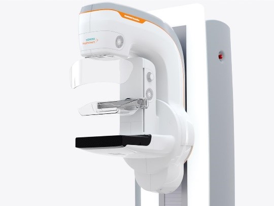
| Parts & Accessories | • X-ray tube • purifier • pressure table • breast table • Network • X-ray detector 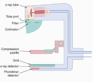 |
| Operation | First, the breast is placed on the designated table. The pressure table moves downward to compress the breast. This procedure is done to flatten the breast as much as possible to obtain a clear image with a low dose of X-ray. A beam of X-rays will be generated from an X-ray tube and passed through the breast to take multiple pictures of each breast (mammogram). - Each photo may take about 20 seconds |
| Common Problems | • A small black spot on the ray angle due to damage to the cassette or exposure of the film to light. In this case, replace the damaged cassette. • White spots on the image due to screen scratches. •Dark marks or excessive brightness due to static electricity. In this case, remove the film from the cassette |
| Manufacturers | • Siemens Healthineers • Philips • GE Healthcare |
| Sources | • Saudi Food and Drug Authority • US Food and Drug Administration • Centers for Disease Control and Prevention • Imaging Department HGS • Radiology Key • Bupa Health UK • MD Anderson Cancer Center • Dentaltix • Siemens Healthineers • Philips • General Electric Healthcare |