
| Properties | Details |
|---|---|
| SFDA Classification | Class IIb |
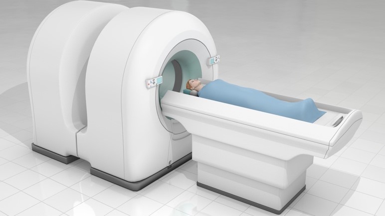
| Purpose of Use | Uses PET Scan Mostly with patients with brain or heart disease and cancer, PET Scan helps It depicts biochemical changes that occur in the body, such as metabolism (the process by which cells convert food into energy after food has been digested and absorbed into the blood) and the heart muscle. |
| Work Location & End-User | Work Location: • Department of Nuclear Medicine. • Department of Oncology. End User: • X-ray specialist. • Nuclear medicine technician. • Radiation therapist. |
| Parts & Accessories | • Mobile patient bed • CT scan room • CT Scanner • Positron emission tomography room Positron emission tomography 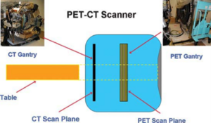 |
| Operation | The principle of positron emission tomography (PET) is that the radiation from radioactive drugs injected intravenously to the patient is recorded by external detectors placed in different directions. The isotope distributes within different tissues according to the carrier molecule (isotope labeling) and emits a positron (positively charged electron). The PET system computer then reconstructs the cross-sectional images from the display data, as does a CT scanner computer. Modern multislice PET scanners allow up to 45 slices to be obtained simultaneously at an axial distance of 16 cm. The spatial resolution of the PET system is limited to 5 mm. PET images are often combined with contrast-enhanced CT images to facilitate anatomical localization of radiopharmaceuticals. |
| Common Problems | • PET Detector Failure • Control Panel Failed Signal processing failed • Data Asylum Failed • Artifacts body imaging • Network problems (transmitting and receiving) • X-ray tube failure |
| Manufacturers | • GE Healthcare • Siemens • Philips • Canon • Toshiba |
| Sources | • Saudi Food and Drug Authority • Johns Hopkins Hospital Medicine • Radiology Key • NCBI • International Atomic Energy Agency (IAEA) |